

Brillouin
Spectrometer
possible outline
The Need
The technology
Description
Listed Accomplishments
Core Services
Optical
We have deep knowledge and expertise in design of development of optical systems for a range of demanding applications.
Some of our Expertise
Microscopy and BioImaging
Precision Laser System Design
Laser Scanning and Modulation
Camera Imaging Systems
Multi-sensor Systems
Lens Design/Simulation (Zemax)
Optomechanical Tolerancing
Stray Light Analysis
Illumination Systems
Mechanical
Mechanical design is integrated into all our products and the design process. We use industry standard computer-aided tools to design individual components, complex systems, and analyze engineering performance.
Some of Our Expertise
CAD (SolidWorks / OnShape)
Precision Opto-mechanics
3D Printing (FDM/SLA)
Electromech Automation/Robotics
Simulation/Analysis
Electrical
Our systems are electronically powered to drive computer automation, light sources, motorized scanners, and peripheral sensors.
Some of Our Expertise
Hardware Architecture
Data Acquisition Systems
Electro-Optical Systems
Chassis Wiring and Management
Circuit Board CAD and Fab
Hardware Automation
Microprocessors and SBCs
Software
We use software to not only control the automation of our electronic devices, but control the presentation and interpretation of data we acquire.
Some of Our Expertise
Algorithm Development
Data/Image Processing
Medical Software Dev
Data Acquisition
Hardware Automation
Graphical User Interfaces
Microprocessors & SBCs

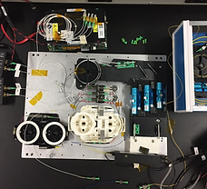
Brillouin Spectrometer
Ophthalmology - Diagnostic/Surgical Planning
Patient specific, high resolution, biomechanical measurement of eye structures is possible using Brillouin Spectroscopy. The technology was spun out of Harvard/MGH to create the first non-invasive laser imaging device for assessing ocular biomechanics.
This complex technology was improved upon (10X signal clarity), drastically reduced in size, multi-modality integration, a modern software interface/database, and automated to be push-button for easy physician use. Several key patents were generated as a result of the development.

Tear Film Interferometer
Ophthalmology - Diagnostic/Research
This system generates movies of the patient's previously invisible tear film through rapid analysis of interferometric fringes. Development was the result of a collaboration with a top optical institution and a fortune 500 Biotech company.
The device was reimagined with optomechanical design for manufacturing, automated 3D motorized alignment, a user friendly software interface, and optimized patient ergonomics. Final units were shipped directly to the client site where clinicians were trained on it use.
OCT Imaging System
Cardiology - Surgical Imaging
Over the course of a 2 year project, developed the Optical Coherence Tomography (OCT) imaging engine for a second generation dual modality imaging catheter system (OCT and Ultrasound). The system acquires high resolution 3D Images of a patient's artery via a specialized fiberoptic catheter in combination with a motorized laser scanning unit. The new generation integrated system was reduced in size and simplified, while achieving superior imaging performance and depth.
The OCT engine was created as part of a larger team effort across multiple organizations to realize the full dual modality imaging system.

Visualization for Vitreoretinal Surgery
Ophthalmology - Surgical Imaging
New drug and delivery mechanisms for the treatment of dry age-related macular degeneration are actively being developed. In this multi-year project, custom imaging/illumination peripherals were developed for ophthalmic surgical microscopes enabling high resolution, wide FOV imaging, and improved safety, during a brand new vitreoretinal surgical procedure.
Prototypes were regularly tested in labs in clinical labs. Microscopes with integrated OCT imaging helped with the understanding of drug delivery within the target tissue.

3D Vision for Robotic Surgery
Surgical Robotics - Stereoscopic Imaging
Real-time dual-HD video processing was developed for management of a next generation single-port laparoscopic robotic gallbladder surgery system.
Interlaced images viewed on a polarized monitor with 3D glasses allowed for lag-free depth perception with overlaid graphics for a seamless surgical robot video interface.
The development was completed as part of a larger program across multiple teams and companies to realize the surgical robotic system.

Laser Scanning Microscopy Systems
Microscopy - Laser Scanning - Laboratory Research
Automated high resolution laser scanning microscopy systems were created from the ground up for tissue engineering research. Laser tweezers, combined with confocal correlation techniques, allowed for sensitive measurement of environmental biomechanics at the scale of individual cells.
A novel high-throughput cell culture device was created and patented for use within developed microscopy systems. This device enabled imaging and mechanical mapping of tissue engineered constructs experiencing gradients of strain in 3D.

Fundus Camera Systems
Ophthalmology - Diagnostic
Through advances in sensors and optics, easy and clear digital imaging of the retina is now feasible using a small handheld device.
We have worked with several optical design forms in different contexts, making technology readily available for stand alone devices as well as integration into larger systems.

Handheld 3D Camera
Dermatology - Stereoscopic Imaging
Dermatology was modernized through creation of this handheld 3D imaging device allowing for high resolution digital tracking of skin moles.
Rapid development of a minimal viable prototype assisted the company's founders to catch another round of Series A funding and market traction.

Dental System Development (Misc)
Dentistry - Prosthetic - Diagnostic
As part of a contracted team, was fortunate to contribute to the design and development of several next generation dental technologies.
These have included:
- Digital impression device which use laser scanning reflection confocal to replace the typical manual bite plate epoxy
- A hand-held impression epoxy dispenser / mixer (patented tip design)
- Bluetooth-enabled dental impact device to detect microfractures within teeth

Gene Therapy Cystic Fibrosis Imaging
Drug Delivery - Clinical Imaging
Cystic Fibrosis is a debilitating genetic pulmonary autoimmune disease in which a missing chloride ion channel protein (CFTR) results in excessive mucus build up in the airways. This published research proved the efficacy of non-viral polymer nanoparticles containing the missing CFTR gene for Cystic Fibrosis in a knock-out animal mouse model.
Multiple imaging modalities (Bioluminescence, Magnetic Resonance Imaging (MRI), and Positron Emission Tomography (PET)) were used in combination to show the clinical efficacy in mice delivered the gene therapy.